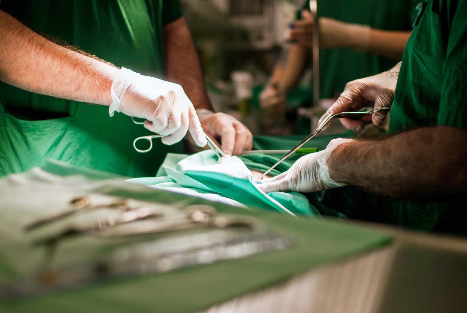
Your patient is a 6-year-old Labrador with a recurrent perineal hernia. The first repair used polypropylene mesh — the standard choice, reasonable cost, familiar technique. Now you’re back in the same field, same defect, and the question isn’t just how to fix it again. It’s whether the material you choose this time will produce a different outcome.
This is where the biological vs. synthetic comparison stops being academic. Every surgeon who has managed a mesh-related complication — seroma, infection, fistula, chronic discomfort at the implant site — has asked whether the material itself was part of the problem. The evidence increasingly suggests that for soft tissue repair in dogs and cats, the answer is yes.
Here’s what the data shows when you put polypropylene and BioSIS ECM side by side.
How Do Polypropylene Mesh and BioSIS Scaffolds Work Differently in the Body?
Polypropylene mesh provides structural support by staying in place permanently. It doesn’t resorb, doesn’t integrate, and doesn’t regenerate — it becomes a chronic foreign body encased in fibrous tissue. The body walls it off rather than incorporating it. That fibrous capsule is how the repair holds, and it’s also the source of the downstream complications: stiffness, contraction, mesh migration, and the inflammatory cascade that never fully resolves because the trigger never leaves.
BioSIS ECM works on a different principle entirely. Porcine small intestinal submucosa is a decellularized extracellular matrix — collagen scaffold, preserved growth factors (VEGF, bFGF), glycosaminoglycans, fibronectin, laminin — that acts as a biological template for the body’s own repair process. Host fibroblasts infiltrate the scaffold, lay down organized collagen, and gradually replace the graft material with native tissue. Studies show cellular infiltration beginning within 2 weeks and tissue resembling native structure by 1 month. By the time remodeling is complete, there is no foreign body — only the patient’s own tissue where the defect used to be.
The practical consequence: polypropylene repairs end with an implant. BioSIS repairs end with tissue.
What Are the Real Complication Rates?
The complication profile of polypropylene mesh in veterinary patients is documented clearly in the literature — and it warrants attention before material selection in any case where infection risk, recurrence, or long-term tissue quality matters.
In a retrospective analysis of perineal herniorrhaphy with cone-shaped polypropylene mesh in dogs, 80.5% of cases were considered successful — but that leaves nearly 1 in 5 dogs with complications within the first 60 days: perineal swelling, persistent tenesmus, and incisional infections. The incisional infection rate was 5.6%, and the recurrence rate reached 12.5%.
More telling is a histological study of polypropylene mesh implanted in dogs via inguinotomy: 100% of animals showed chronic foreign body reaction in the surrounding tissue. The inflammatory cell volume in polypropylene mesh repairs was 32% — compared to 8% in polyester and 12% in ePTFE. That chronic inflammatory burden isn’t a complication in the traditional sense. It’s the expected tissue response to a permanent implant.
Seroma formation, which can persist for weeks to months post-operatively, is a common sequela in both synthetic and biological repairs, though its significance differs: with synthetic mesh, a seroma can become a track for contamination around an implant that can’t be removed without reoperation. With BioSIS, fluid accumulation typically resolves as the scaffold remodels and native tissue replaces it.
Fistula and mesh erosion — particularly in perineal locations with proximity to bowel — are rare but well-documented complications of permanent synthetic implants. There is no equivalent with biological scaffolds, which are fully resorbed before tissue remodeling is complete.
Why Does Scar Tissue Form With Synthetic Mesh—and Not With BioSIS?
Scar tissue is the body’s default response to an unresolvable injury signal. With polypropylene mesh, that signal doesn’t resolve — the implant is permanent, and so is the low-grade inflammatory response surrounding it. Macrophages activate, fibroblasts deposit dense fibrous tissue around the mesh, and what forms is not organized native tissue but a fibrous capsule encasing a foreign material. Over time, that capsule contracts, which is one mechanism behind the chronic discomfort and reduced tissue pliability seen in long-term synthetic mesh repairs.
BioSIS short-circuits this pathway. Because the scaffold is gradually degraded and replaced — not walled off — the macrophage response shifts from pro-inflammatory to pro-remodeling. The preserved growth factors in the ECM (VEGF and bFGF in particular) support angiogenesis and cell proliferation rather than driving a chronic foreign body reaction. A 2025 meta-analysis of SIS-ECM composition confirmed these preserved bioactive factors as central to the regenerative mechanism that distinguishes biological scaffolds from synthetic materials.
The clinical result: BioSIS repairs heal with organized, vascularized tissue. Polypropylene repairs heal with fibrous encapsulation around a permanent implant. The distinction matters most in high-mobility areas — perineal, abdominal wall, diaphragmatic — where tissue quality and pliability affect long-term function.
When Should You Choose BioSIS Over Synthetic Mesh?
The honest answer is that BioSIS is appropriate for a broader range of cases than it’s currently used in. But the clinical scenarios where it has a clear advantage over polypropylene are worth knowing specifically.
Contaminated or potentially contaminated fields. Polypropylene mesh in a contaminated wound carries substantial risk — biofilm formation on a permanent implant can require complete mesh removal, a significantly more complex second surgery. BioSIS in a contaminated field does not carry the same risk of harboring persistent infection because it is resorbed as it heals.
Pediatric and growing patients. A permanent implant placed in a young, growing patient creates a structural constraint as surrounding tissue develops. A resorbing scaffold does not — the repair integrates with native tissue that can grow and remodel normally.
Recurrent hernias after prior synthetic repair. As in the Labrador scenario above: after a failed polypropylene repair, the tissue environment is already compromised by prior foreign body reaction and fibrosis. BioSIS offers a path to organized regeneration rather than layering another permanent implant into a scarred field.
Peritoneal contact risk. Polypropylene mesh in contact with bowel carries risk of adhesion formation and erosion over time. Biological scaffolds are significantly safer in anatomic locations where intraperitoneal placement is necessary.
When tissue quality is the primary concern. For owners who want their animal’s long-term outcome to reflect organized, native tissue repair — not a permanently implanted foreign material — BioSIS delivers that outcome. That’s a conversation worth having during surgical planning, particularly for active working dogs or high-expectation clientele.
A 2024 case report documented successful use of porcine SIS to repair a congenital diaphragmatic hernia in a kitten — a structural repair in a critical anatomic location in a pediatric patient, with excellent outcome. It illustrates both the versatility of biological scaffolds and the clinical situations where the regenerative mechanism is most valuable.
The Bottom Line for Material Selection
Polypropylene mesh is not the wrong choice for every patient. It’s cost-effective, technically familiar, and appropriate where the risks of chronic foreign body reaction are acceptable. But the evidence is clear that its permanent implant nature carries a chronic inflammatory burden — 100% foreign body reaction in every patient, documented in the veterinary literature — and that complication risk is real, not theoretical.
BioSIS ECM offers a different outcome: complete resorption, organized tissue regeneration, no permanent foreign body, and a complication profile driven by patient biology rather than implant persistence. For cases involving contamination risk, recurrence, pediatric patients, or peritoneal contact, the biological scaffold isn’t just an alternative — it’s the better choice.
For more on how BioSIS ECM is used across soft tissue repair cases in dogs and cats, visit RethinkHealing.com.
 According to the
  Your furbaby needs gastrointestinal surgery, and you have all kinds of questions. We’re here to discuss how you can prepare and what you need to know.