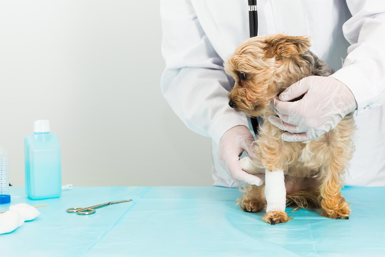
It’s day 18. The wound is clean, it’s moist, it’s covered. And it looks exactly the same as it did at day 10. No new granulation islands. No edge contraction. The exudate has shifted from serous to something slightly cloudier. The tissue around the margin is pale instead of the pink-red you’d expect in an actively healing wound.

This wound has stalled — and continuing the current protocol isn’t going to change that.

Chronic non-healing wounds are one of the most frustrating management problems in small animal practice. Not because they’re rare, but because standard wound care is genuinely appropriate for most wounds — and the moment when it stops being appropriate isn’t always obvious. Understanding what biologically derails the healing cascade, which patients are predictably at risk, and what a biologic intervention actually does differently is what separates a wound that closes in five weeks from one that doesn’t close in five months.

The Biology of the Stalled Wound: What Goes Wrong When Healing Stops

Normal wound healing follows a defined sequence: hemostasis, inflammation, proliferation, remodeling. Each phase has a beginning and an end. In a stalled wound, the sequence breaks down at the transition from inflammation to proliferation — and the wound gets stuck cycling through an inflammatory response that can’t resolve.

Three mechanisms drive most of what goes wrong.

Biofilm. Biofilm is present in approximately 60% of chronic wounds, compared to just 6% of acute wounds. That difference isn’t incidental — biofilm is both a consequence of chronicity and a driver of it. The extracellular polymeric substance (EPS) matrix that holds biofilm together creates three compounding barriers: a physical barrier that blocks keratinocyte and fibroblast migration; a metabolic barrier, generating hypoxic and acidic gradients that suppress cell energy production; and an immunological barrier that masks bacterial surface signals from host immune cells while bacterial toxins actively damage wound bed tissue. S. aureus α-toxin depletes ATP in keratinocytes. P. aeruginosa phospholipase causes direct necrosis. The biofilm doesn’t just prevent healing — it actively dismantles the wound bed.

MMP dysregulation. Matrix metalloproteinases are necessary for normal wound remodeling, but in chronic wounds, elevated MMP-9 creates a protease-dominant environment that degrades the extracellular matrix scaffolding before new tissue can form. Bacterial proteases compound this — S. aureus degrades immunoglobulins and complement proteins; certain anaerobes target type IV and V collagen directly. The wound bed loses its structural matrix faster than it can replace it.

Cell cycle arrest. Fibroblasts from chronic wounds exhibit growth arrest driven by elevated CDK inhibitors p21 and p27. These cells accumulate in the wound but can’t proliferate. Meanwhile, excess TGF-β — present in chronically inflamed wounds — imposes a secondary brake on keratinocytes via Smad2/3 phosphorylation, preventing the epithelial migration needed to close the wound surface. The cellular machinery is present but biochemically locked.

The result is a wound that looks stable but is doing nothing — consuming resources, resisting standard dressings, and waiting for a different signal.

Which Patients Get Stuck: Common Causes in Dogs and Cats

Certain wound types and patient profiles are predictably prone to chronicity. Recognizing them early changes the management calculus.

Post-surgical dehiscence. The most common scenario in general practice. A wound that should have closed primarily reopens — most often due to tension, infection, or patient interference — and then fails to granulate adequately over the following weeks. The surgical field has already been traumatized once; fibroblasts in the wound margins may be compromised, and secondary biofilm establishment is common. Revision surgery in a poorly vascularized, previously disrupted field carries its own risks. This is often where a biologic intervention has its clearest return.

Pressure ulcers in recumbent patients. Dogs with neurological or orthopedic conditions who are non-ambulatory develop pressure sores over bony prominences that are mechanically and metabolically difficult to heal. Sustained pressure impairs capillary perfusion, creating a hypoxic wound bed — precisely the environment that prevents normal cellular proliferation and is most permissive to biofilm. These wounds can persist for months to years under standard care.

Bite wounds and infected wounds in immunocompromised patients. FIV-positive cats, patients on chronic corticosteroids, and animals with concurrent systemic disease represent a high-risk group. A bite wound in a healthy cat is an acute problem with a predictable course. The same wound in an immunocompromised patient can fail to resolve for months, particularly when polymicrobial antibiotic-resistant biofilm establishes in the wound bed.

Patients with endocrine disease. Hyperadrenocorticism and diabetes mellitus are well-documented drivers of impaired wound healing in dogs. Elevated cortisol suppresses fibroblast function, impairs collagen synthesis, and blunts the immune response needed to clear early wound contamination. Any patient presenting with a chronically non-healing wound that doesn’t fit the wound’s timeline should be screened for underlying endocrinopathy — no biologic intervention fully compensates for uncontrolled Cushing’s disease.

The common thread across these patients is that their wound biology has been disrupted at a systemic or local level in a way that standard topical care cannot address.

How Amniotic Membrane Powder Restarts the Cascade — Step by Step

Standard wound care — appropriate moisture, debridement, infection control — creates the conditions for healing. What it doesn’t do is provide the biochemical signal the wound needs to move from inflammation to proliferation. That’s the gap a biologic like Healion fills.

Decellularized amniotic membrane powder delivers a preserved payload of growth factors that are absent or depleted in the chronic wound environment. Measured concentrations from amniotic membrane preparations include epidermal growth factor (EGF), fibroblast growth factor (FGF), hepatocyte growth factor (HGF), keratinocyte growth factor (KGF), nerve growth factor (NGF), and TGF-β — the same signals that coordinate normal wound healing but that chronic wounds can no longer generate at adequate levels.

Lyophilized amniotic membrane preparations show significantly higher bioactive factor concentrations than fresh tissue — up to 3.8x the collagen content, 5.2x the VEGF, and 6.8x the bFGF per milligram. That concentration matters clinically: a lyophilized powder applied to a chronic wound bed delivers a more concentrated signal than fresh membrane or simple moist dressings.

The mechanism at the cellular level is well-characterized. In chronic wounds, excess TGF-β locks keratinocytes in a growth-arrested state via Smad2/3 phosphorylation — the cells can’t migrate or proliferate. Amniotic membrane EGF activates MEK and attenuates Smad2/3 phosphorylation, releasing the TGF-β-imposed brake on keratinocyte proliferation. This isn’t blunt inflammation suppression — it’s modulation of a specific pathway that the chronic wound has dysregulated. Minimal TGF-β signaling is still necessary for proper migration; the AM restores balance rather than eliminating a signal.

Simultaneously, amniotic membrane shifts macrophage behavior from pro-inflammatory (M1) toward pro-remodeling (M2), elevating IL-10 (anti-inflammatory), supporting controlled TGF-β for appropriate matrix remodeling, and increasing MMP-1 and MMP-3 for organized ECM turnover. The net effect is a wound environment that can finally transition out of the inflammatory phase and begin building new tissue.

In a controlled study of lyophilized amniotic membrane in dogs with full-thickness wounds, closure rates at week 3 were 88.6% in the treatment group versus 55.7% in controls. By week 5, treatment wounds showed restored epidermis, remodeled dermis, and recovered hair follicles — structural markers of genuine tissue regeneration, not just wound closure. A separate AVMA study of an ECM wound dressing in Beagles showed significantly higher epithelialization percentages at days 7, 9, 12, and 18 compared to standard protocol, with better histologic repair scores.

Healion also retains antimicrobial activity from native amniotic tissue — β-defensins with documented activity against P. aeruginosa and S. aureus including MRSA strains. In a wound where biofilm is part of the problem, this is a clinically meaningful addition to the protocol.

Practical Decision Points: When Should a GP Vet Reach for a Biologic?

The clinical question isn’t whether amniotic membrane powder works — the evidence supports it. The question is when in the management timeline it becomes the right tool.

A reasonable framework: if a wound is not showing clear signs of progression by day 14 to 21 under appropriate standard care, treat that as a stall signal rather than normal variation. Specifically:

  • No granulation islands forming at 14 days
  • Wound edges not contracting
  • Exudate shifting from serous to cloudy or purulent despite appropriate antibiotic coverage
  • Tissue at the wound margin appearing pale or grey rather than pink-red
  • Culture showing resistant organisms or polymicrobial contamination

At that point, consider whether there’s a systemic reason the wound is stalling — run a culture if you haven’t, screen for endocrine disease if the patient profile suggests it, reassess nutrition and pain management. Then layer in the biologic.

Healion’s bellow dispenser allows direct, sterile application to the wound bed without secondary processing — the powder format suits open wounds with irregular geometry and doesn’t require the wound to be prepared beyond standard debridement. It fits into an existing wound care protocol without requiring additional equipment or technique change.

The conversation with the client is straightforward: standard dressings keep the wound clean and provide the right environment. The powder provides the biological signal the wound has lost — growth factors and structural cues that tell the cells what to do next. That framing holds up to client questions and positions the intervention accurately.

For patients at high risk of chronicity — post-surgical dehiscence in an oncology patient, pressure sores in a recumbent dog, bite wounds in an immunocompromised cat — there’s a reasonable argument for starting the biologic earlier rather than waiting for the stall to establish.

Learn more about Healion’s mechanism and application at RethinkHealing.com.

A client returns for a two-week post-op recheck after a routine mass removal on their dog’s flank. The incision is clean, the dog is comfortable, and healing is progressing normally. Then the client asks the question you’ll hear again and again: “Is the scar going to look like this forever?”

The honest answer is more nuanced than most vets have time to explain in a recheck appointment. Scar formation is a biological process that begins in the first hours after wound closure and is largely determined within the first three weeks — long before the scar is visible enough for an owner to judge. By the time a client notices a raised, thickened, or discolored scar at week six, the biology that created it is already finished.

That window — the acute inflammatory phase — is where intervention matters. And it’s where amniotic membrane powder changes what’s clinically possible.

Why Wounds Scar: The Three-Week Window That Matters

Normal wound healing proceeds in three overlapping phases: inflammation, proliferation, and remodeling. Scar tissue is the product of what happens in the transition between inflammation and proliferation — specifically, how well the wound resolves its inflammatory state before fibroblasts begin depositing collagen.

The key driver is TGF-β (transforming growth factor beta). Released by platelets and inflammatory cells immediately after injury, TGF-β recruits fibroblasts to the wound and signals them to produce collagen. This is necessary for wound closure. The problem arises when TGF-β signaling persists too long or is too intense — fibroblasts overproduce collagen, depositing it in disorganized bundles rather than the aligned architecture of normal dermis. The result is scar tissue.

Critically, research has shown that TGF-β suppression works at the transcriptional level — amniotic membrane downregulates the gene expression of TGF-β2, TGF-β3, and all three TGF-β receptor types in fibroblasts. This isn’t a surface-level effect. It’s an upstream interruption of the signaling cascade that determines whether a wound heals cleanly or scars.

The window for this intervention is weeks one through three. After that, the collagen framework is laid and the remodeling phase determines final scar quality — a slower, less modifiable process. Scars typically appear most pronounced at three to eight weeks post-closure and reach final form at six to twelve months. What they look like at week six was determined biologically in week two.

Can You Actually Prevent Scarring in Post-Surgical Wounds?

The short answer is: not completely, but meaningfully — if the intervention targets the right mechanism at the right time.

Standard post-op care focuses on infection prevention, moisture management, and keeping the patient from disrupting the wound. These are essential. But none of them address the inflammatory signaling driving fibrosis. Suturing technique affects scar width and tension. Infection increases inflammatory load. But the underlying TGF-β cascade runs regardless of how clean the incision is or how well the edges are apposed.

Amniotic membrane works through a distinct pathway. It modulates the macrophage response — shifting the population away from the pro-inflammatory M1 phenotype that amplifies TGF-β signaling, toward the pro-resolving M2 phenotype that supports organized tissue remodeling. This shift matters because macrophage polarization in the first one to two weeks is one of the strongest predictors of scar outcome in healing wounds.

A 2025 study published in Scientific Reports evaluated lyophilized (powdered) amniotic membrane on full-thickness skin wounds in dogs and found efficacy comparable to fresh membrane — validating that the decellularization and powdering process preserves the biologically active components responsible for healing modulation. Studies on canine amniotic-derived products have consistently demonstrated benefits in both in vitro models and in vivo wound healing, with outcomes including accelerated epithelialization and reduced inflammatory burden.

The practical implication: applying amniotic membrane powder during the acute phase — at closure and at early rechecks — gives the wound biology a better trajectory before the collagen deposition phase begins.

Why Cosmetic Outcomes Matter More Than Vets Often Assume

Post-surgical scar appearance is not a vanity concern — it’s a measurable driver of client satisfaction and practice loyalty. A study published in Veterinary Evidence found that cosmetic outcome as judged by the owner accounted for 37% of the variability in overall satisfaction with surgical procedures in dogs. That number is significant: roughly one in three points of client satisfaction after surgery is determined by how the wound looks, not how quickly the patient recovered or how well the procedure went.

There is also documented discordance between how veterinarians and owners assess incision appearance. Vets evaluate healing on clinical criteria — no dehiscence, no infection, appropriate healing progression. Owners evaluate on cosmetic criteria — hair regrowth, scar size, color, texture. These are different standards, and vets who address only the clinical criteria miss the metric that determines whether a client returns.

This matters particularly for procedures in cosmetically visible locations: facial mass removals, limb surgeries, flank incisions on short-coated breeds, and any procedure where hair regrowth may be incomplete. For breeders, show animals, or clients who simply place high value on appearance, the post-surgical cosmetic outcome is often the lasting impression of the entire clinical experience.

Adding Scar Prevention to Your Post-Op Protocol with Healion

Healion delivers decellularized amniotic membrane in a powder format via a sterile bellow dispenser — a practical design for post-surgical wound application where liquid or sheet formats would require more handling or risk of contamination.

The protocol addition is straightforward:

  • At closure — apply Healion to the wound bed before subcuticular or skin closure in cases where scar risk is elevated (cosmetically sensitive location, large wound surface area, high-tension closure, or breed predisposition to scarring)
  • Early post-op rechecks (days 3–7) — apply to the incision line if any wound edge separation, inflammation, or slow re-epithelialization is noted
  • Acute lacerations and traumatic wounds — apply after debridement to contaminated wounds where scar formation is expected given the wound mechanism

Healion modulates inflammation, enhances healing, and reduces scar tissue formation — beginning at the cellular level in the first week before scar architecture is established. The bellow dispenser allows precise, hands-off application directly to the wound surface without the mixing or handling challenges of liquid biologics.

Setting Client Expectations After Application

When you add Healion to a post-surgical protocol, give clients a clear timeline: “The wound will still go through a visible healing phase over the next several weeks. What we’re doing now is giving the tissue the best biological environment to heal with less scar formation. You’ll be able to judge the final result at about six months.” That framing manages expectations while communicating that you took a proactive step most practices don’t offer.

Clinical Bottom Line

Scar formation is determined in the first three weeks of healing — not at the six-week recheck when clients first comment on appearance. The biology is settled before the scar is visible. Intervening during the acute inflammatory phase, when TGF-β signaling and macrophage polarization are still modifiable, is the only window for meaningful scar prevention.

Healion brings an evidence-supported mechanism to that window: amniotic membrane matrix that suppresses fibrotic signaling, modulates inflammation toward resolution, and supports organized tissue remodeling from the first application. For surgical practices where client satisfaction matters — and where cosmetic outcome accounts for more of that satisfaction than most vets realize — it’s a meaningful addition to the post-op toolkit.

Explore Healion’s clinical profile and request a sample at RethinkHealing.com.

A 6-year-old mixed-breed dog presents three weeks post-degloving injury. The wound is clean. Granulation tissue started, then stalled. You’ve cycled through wet-to-dry bandages, silver dressings, and a course of systemic antibiotics. The tissue looks flat. No measurable progress. The owner is frustrated and asking what comes next.

This is the chronic wound scenario that sends many practitioners back to the textbook. And it’s exactly the situation decellularized amniotic membrane powder was developed to address.

Healion, Vetrix’s veterinary amniotic wound powder, delivers a concentrated source of bioactive matrix proteins, growth factors, and anti-inflammatory signaling molecules directly to the wound bed. Here’s what the science says — and when to add it to your wound care toolkit.

Why Chronic Wounds Stall — and Why Standard Care Can’t Fix It

Acute wounds heal in a predictable sequence: hemostasis, inflammation, proliferation, remodeling. Chronic wounds don’t follow that sequence. They cycle through inflammation without resolving, accumulate senescent fibroblasts, and fail to transition into the proliferative phase that drives closure.

Repeated bandage changes, antimicrobial dressings, and topical antibiotics address symptoms. They don’t reset the signaling environment. Wound fluid in chronic cases is biochemically different from acute wound fluid — it’s rich in matrix metalloproteinases (MMPs) that degrade growth factors before they can act, and depleted of the cytokines that recruit reparative cells.

Standard moist wound healing (MWH) remains the foundation of veterinary wound management. But for wounds stuck in chronic inflammation, moisture retention alone isn’t sufficient. Something needs to shift the cellular environment — not just cover it.

That’s where amniotic membrane biologics enter the picture. The mechanism isn’t a newer dressing or a better antiseptic. It’s a fundamentally different approach: restoring the biological signals the wound needs to move forward.

How Decellularized Amniotic Membrane Powder Works

Amniotic tissue is uniquely anti-inflammatory by origin. It forms at the maternal-fetal interface — an immunologically privileged environment where the body must suppress rejection while simultaneously supporting rapid new tissue formation. That immunomodulatory capacity is preserved when amniotic membrane is decellularized and processed into powder form.

Three mechanisms are well-documented in peer-reviewed literature:

Macrophage reprogramming. Amniotic membrane traps pro-inflammatory neutrophils and M1 macrophages and induces apoptosis. At the same time, it skews macrophage differentiation toward the M2 phenotype — the reparative subtype that drives angiogenesis, fibroblast recruitment, and re-epithelialization. In a chronic wound, this shift is often the step that’s missing entirely.

TGF-β suppression. TGF-β (transforming growth factor beta) is the primary driver of fibrosis. In chronic wounds, sustained TGF-β signaling doesn’t produce organized tissue — it produces scar. Amniotic membrane suppresses this pathway, protecting the wound from excessive scar tissue formation. This is the mechanism behind Healion’s documented ability to reduce scarring, an outcome clients often notice and appreciate — particularly in cosmetically sensitive areas like the face or limbs.

Growth factor delivery. Decellularized amniotic matrix retains a preserved scaffold of HGF (hepatocyte growth factor), EGF (epidermal growth factor), FGF (fibroblast growth factor), and PDGF (platelet-derived growth factor). These proteins drive keratinocyte migration, fibroblast proliferation, and vascular ingrowth — the cellular machinery of closure.

A 2025 systematic review published in Springer Nature confirmed that amniotic membrane transplantation reduces inflammatory cytokines and inflammatory cell density in chronic wound biopsies compared to controls. In a canine clinical study, wounds treated with amniotic-derived conditioned media showed a 98.47% reduction in wound surface area at 15 days — compared to 57.14% in the control group — with complete histological regeneration by six weeks. (Source: International Journal of Molecular Sciences, MDPI)

The powder format is clinically significant. Unlike liquid biologics or hydrogel formulations that require mixing or direct manual contact during application, Healion’s bellow dispenser delivers sterile powder directly to the wound bed without introducing handling contamination. In wounds with irregular geometry — undermined edges, exposed tissue pockets, uneven granulation — powder conforms to the wound surface in ways a sheet or hydrogel cannot.

Can You Use Healion on Contaminated or Infected Wounds?

This is the question most practitioners ask first. The short answer: prepare the wound bed before applying.

Healion is not an antimicrobial product. However, amniotic membrane does express antimicrobial peptides — research on canine amniotic tissue has identified β-defensins 1 and 103 and Elafin, which show documented activity against Pseudomonas aeruginosa and Staphylococcus aureus, including MRSA strains. This provides a degree of protection against secondary contamination once the wound is prepared, but it does not substitute for active infection management.

The recommended clinical protocol:

  • Debride to viable, well-vascularized tissue
  • Culture if infection is suspected or the wound has been chronic
  • Resolve active infection with appropriate systemic or topical antimicrobials
  • Apply Healion once the wound bed is clean — granulating or stalled pre-granulation

This sequence matters. Applying a biologic to an infected wound bed doesn’t accelerate healing — the inflammatory burden from active infection will overwhelm the signaling benefit. The goal is to introduce Healion into a wound that’s clean but biologically stuck.

Once applied, re-bandage with a non-adherent primary layer. Standard secondary and tertiary bandage layers apply. Recheck timing depends on wound type and patient compliance, but most chronic wound cases benefit from reassessment at 7–10 days to document progress before the next application decision.

When to Reach for Healion: Clinical Indications and Patient Selection

Healion is indicated where the healing cascade has stalled and conventional management is no longer producing results. Practical trigger points:

  • Chronic wounds >3–4 weeks without measurable closure progress
  • Degloving injuries with large surface area or irregular wound margins
  • Post-surgical dehiscence when primary or delayed closure isn’t viable
  • Pressure wounds and decubital ulcers in debilitated or recumbent patients
  • Traumatic wounds with exposed deep structures as an adjunct to reconstructive planning
  • Cosmetically sensitive locations where scar reduction is a priority — face, limbs, areas of expected hair regrowth

Healion is not a first-line treatment for acute wounds that are progressing on standard protocol. Those wounds heal well with conventional care. Healion is the intervention for cases where you’re three or four weeks in, the wound looks clean, and nothing is moving.

Patient selection is straightforward. Any dog or cat with a nonhealing wound and a prepared wound bed is a candidate. Decellularized amniotic membrane is immunologically inert by structure — the decellularization process removes cellular components that trigger immune response, leaving behind the extracellular matrix scaffold. No documented biocompatibility concerns exist for canine or feline patients.

What Clients Will Notice

For wounds that have plateaued for weeks, client communication is as important as clinical management. When healing accelerates after Healion application, clients see the change — and it shifts their confidence in the treatment plan.

Reduced scar tissue formation is often the outcome clients comment on most, especially in dogs with wounds on the face or legs where cosmetic outcome matters. Setting clear expectations — “we’re introducing a biologic that changes how the tissue signals at the cellular level, not just a different dressing” — helps clients understand why this step is different from everything that’s been tried so far.

Clinical Bottom Line

Chronic wounds are one of the most frustrating problems in small animal practice — not because the diagnosis is unclear, but because standard treatment reaches its ceiling and progress stops. The biology behind that stall is well-understood: the signaling environment is disrupted, and no dressing change will fix it.

Decellularized amniotic membrane powder addresses that directly. Healion modulates inflammation, suppresses fibrosis, and delivers the growth factor signals the wound needs to move forward — in a sterile, easy-to-apply format built for veterinary wound management.

For the cases on your treatment board that aren’t moving, it belongs in the conversation. Explore Healion’s clinical profile and request a sample for your practice at RethinkHealing.com.

Revolutionizing Veterinary Wound Care with Healion: A Case Study in Regenerative Medicine

In the ever-evolving field of veterinary medicine, innovative products like Healion Amniotic Wound Matrix are setting new standards for wound healing. By utilizing the powerful properties of amnion, Healion provides veterinarians with a reliable and effective tool for treating complex wounds, enhancing the natural healing process. This blog post will explore the remarkable healing journey of a turtle, showcasing the benefits of Healion in veterinary wound care.

The Role of Amnion in Regenerative Medicine

Amnion, the innermost layer of the placenta, is packed with growth factors, anti-inflammatory properties, and essential proteins that are crucial in wound healing. The Healion Amniotic Wound Matrix, derived from this unique tissue, is delivered in a powdered form, making it easy to apply. This product promotes rapid tissue regeneration, reduces inflammation, and provides a bioscaffold for new cells to grow, making it an invaluable solution in regenerative medicine.

Case Study: A Turtle’s Fast-Track to Recovery

Dr. Garry Innocent, a veterinarian at All Species 24hr Clinic in Alpharetta, GA, encountered an exotic patient with a severe head injury. The turtle had been scalped, leaving no tissue to facilitate traditional wound closure. Faced with this daunting challenge, Dr. Innocent turned to Healion Amniotic Wound Matrix.

Within 24-36 hours of applying Healion, the results were nothing short of extraordinary. The wound began healing rapidly, showing organized, healthy tissue growth. Dr. Innocent expressed his satisfaction, noting that Healion was simple to apply and did not lead to further complications, unlike traditional wound treatments which sometimes cause additional trauma.

For a case where minimal tissue was available and the delicate nature of the patient complicated suturing, Healion’s trauma-free application was the perfect solution. The turtle’s speedy recovery demonstrates the effectiveness of this regenerative wound care tool across different species.

Why Healion Is Essential for Veterinarians

Healion’s amnion-based matrix provides a safe, versatile, and easy-to-use solution for a wide range of wound types, including surgical wounds, trauma injuries, and ulcers. Its unique properties not only accelerate healing but also minimize scarring, making it a preferred choice in complex cases where traditional methods fall short.

The ability to promote natural tissue regeneration without causing additional trauma makes Healion indispensable for treating patients where suturing isn’t viable or safe. Additionally, its application is simple, reducing the stress for both veterinarian and patient, whether treating small, exotic animals or common household pets.

Conclusion

Healion Amniotic Wound Matrix is revolutionizing the way veterinarians approach wound healing. By leveraging the regenerative properties of amnion, it offers an effective solution for complex wounds that are difficult to close or heal. In the case of Dr. Innocent’s turtle, Healion proved its ability to facilitate fast, trauma-free healing in a delicate patient, underscoring its versatility and power as a regenerative medicine product. For veterinarians seeking reliable and advanced wound care options, Healion is a must-have in the medical toolkit.

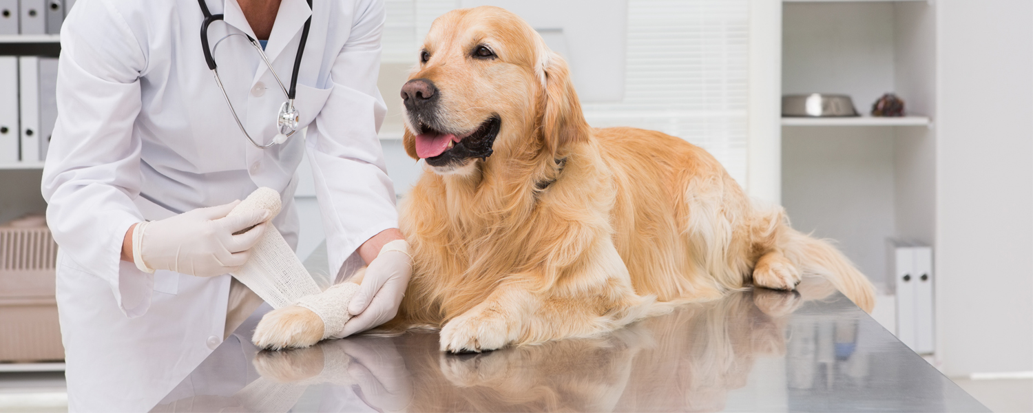
Small dog on a vet’s tableBite wounds are one of the most common reasons dogs see their veterinarians. If a dog is bitten or gets into a fight with another dog, this is what needs to happen first:
  1. As the dog’s owner, stay calm. Panic will only make the situation worse.
  2. Don’t get between your dog and the other dog(s) to break up the fight. You could also get bit in the process.
  3. Focus on getting your dog away from the other dog. A loud clap or another distracting sound may help get your dog’s attention so you can call or signal him to you.
  4. Don’t scream at the other dog, as this could make the situation feel even more threatening to the dogs.
  5. Ask the other dog owner (if present) if their dog is up to date on their vaccines. If the pet owner isn’t present or the other dog involved is a stray, try to get pictures at the very least.
  6. Once your dog is safely away from the dog who bit him, contact your vet immediately or head to your nearest emergency animal hospital.

Why Do Dogs Bite?

Biting is often part of play for puppies. In adult dogs, multiple motives or causes for biting can stem from competitive issues or reactions to perceived threats. Additionally, several factors influence the severity of a dog bite and the health risk it can pose to a pet. This is why seeing your veterinarian as quickly as possible after your dog has experienced a bite is a good idea.

Treating Bite Wounds in Dogs

Dog bites can cause significant injury to the skin and soft tissues. A dog’s teeth and jaws are powerful; a bite wound can result in torn muscles and skin. Not all bite wounds are big, though. Just because a bite wound is small doesn’t mean it isn’t a cause for concern. Minor bites can heal themselves quickly but, in doing so, can trap bacteria in the wound that can result in an abscess.

Infections are the primary concern for any dog bite, but other serious issues can develop if not treated properly. Typically, a vet will examine and thoroughly clean the bite wound and then provide a broad-spectrum antibiotic to help fight infection and prevent further complications with the wound site. Or, the vet can use Vetrix’s Healion Amniotic Wound Matrix as part of their patient’s bite wound treatment plan. 

Healion is a decellularized amniotic membrane supplied in fine, dry, sterilized white powder form for veterinary use. Healion amniotic wound matrix leverages the natural healing properties of the amnion to modulate inflammation and create an environment conducive to rapid cell migration.

You apply it to the wound site and appropriately bandage it to create a sterile healing environment. The bite should be re-examined every 4-5 days, and a single application is usually sufficient.

How To Help A Dog Heal After A Bite

As a vet, your priority is preventing your patient’s bite wound from getting infected. Start treating bite wounds with a fast and effective treatment option that makes patients and clients happy. Add Healion to your medical toolkit today. Order now.

BioSIS wound dressing photolarge copyAny veterinarian worth their salt knows that providing superior wound care treatment starts with assessing the overall stability of the animal. Once the patient is stable, the next step is administering first aid for the wound as soon as safely possible. Your treatment should follow the fundamentals of debridement, infection or inflammation control, and moisture balance. Success depends on taking the correct approach and deciding whether to manage the wound as open or closed.
Before considering surgery, veterinarians looking for a more effective wound treatment should explore Healion Amniotic Matrix. The solution is an easy-to-use, sterilized powder that promotes the healing of wound sites.

Provide Superior Wound Care with Healion Wound Matrix

When choosing wound care solutions for your patients, protecting the wound sight from further contamination and trauma is often a top concern. This is where Healion Wound Matrix can help, especially in situations where a wound cannot or should not be closed. Healion is a decellularized amniotic membrane supplied in fine, dry, sterilized white powder form for veterinary use. It leverages the natural healing properties of amnion to modulate inflammation and create an environment conducive to rapid cell migration. All you must do is puff the solution onto the wound bed and cover it with a non-adherent bandage.

Once administered, and as we touched on at the beginning of this post, it’s advised you consider the following four things while caring for your wound patient:

  1. Giving the wound proper support
  2. Maintaining proper moisture levels
  3. Mitigating infection risk
  4. The phases of wound healing

If these four factors aren’t considered when bandaging and monitoring your patient, you might not see the healing you expect. (Remember pain management throughout the phases of recovery, too.)

Healion Wound Matrix serves as a wound care solution where open wound management or delayed wound closure is the best course of action for the patient. Healion is uniquely designed to enable veterinarians to provide superior wound care treatment and is indicated for the management of wounds, including:


  • Partial wounds
  • Full-thickness wounds
  • Surgical wounds
  • Trauma wounds
  • Draining wounds

Do You Want to Provide Superior Wound Care Treatment?

It’s time to join top veterinarians using the best regenerative wound care technology and provide your patients with faster and more effective treatment options. Healion Wound Matrix provides an ideal healing environment and flexibility in treating both acute and chronic wounds, and it’s affordable.

Healion is cost-effective and packaged to fit procedural needs. Learn about Healion Wound Matrix pricing (available in 6-pack, 12-pack, and 24-pack options).

Veterinary Neurology Vetrix

When a patient has a wound that’s too big to close on its own or a wound that becomes chronic and doesn’t heal by itself, treatment with a skin graft may be beneficial in obtaining wound closure. Skin grafts help quickly and effectively restore function and provide better cosmetic results than other treatment options. Additionally, skin grafts protect from infections and parasites. The Vetrix BioSIS Technology we provide is a regenerative medicine beneficial in remodeling, regrowing, and repairing wounds encountered in the veterinary field.

Two Common Skin Graft Procedures in Veterinary Medicine

When it comes to common household pets, veterinarians are often most likely to perform one of the following types of skin grafts when necessary for wound treatment:

  • Total Skin Graft (or Full-thickness skin graft): This skin graft includes the epidermis and dermis. It involves removing a piece of skin and the fat from the underside of the skin. This grafting procedure requires the donor site to have enough surrounding loose skin so the incision can be closed.
  • Partial Thickness Skin Graft (or Split-thickness skin graft): This skin graft involves shaving a thin layer of skin, 0.2 to 0.4mm, off the donor site. The cutting plane of this graft remains above the hair follicles so that no hair will grow from this skin graft. The donor site will heal independently and doesn’t require closure, but it may be more painful than a total skin graft because the exposed nerve endings will need time to heal. It’s typically used in burn victims with limited normal skin for grafting.

Veterinarians will guide their patients on the best skin graft option for their particular wound.

Treating Skin Grafts With Regenerative Veterinary Medicine

Once it’s been determined that a pet needs a skin graft to heal a wound, consider a regenerative medicine solution as part of your treatment. Unlike synthetic materials or other biological grafts, Vetrix Technology provides a structure for healthy tissue to grow across and incorporate into the extracellular matrix, resulting in wholly remodeled, vital, and fully vascularized tissues. During the healing process, Vetrix BioSIS is replaced with the body’s native tissue, developing into a permanent repair without the long-term presence of a foreign body. After healing is complete, patients are left with healthy, natural tissue.

BioSIS is a great regenerative medicine solution for skin grafts because it facilitates angiogenesis and rapid remodeling, allowing the body’s defense mechanisms to react and respond to any potential infection. It promotes safe and sterile healing of the wound.

Download the BioSIS Informational Brochure to learn more about this technology and how it can aid in healing skin graft patients.

Get the brochure.

We know you want to give your patients the best care there is. From diet and nutrition to coat care to wound care, you want to feel confident that you’re up to speed on the latest technology and products. This can be difficult when there’s so much information out there. When it comes to wound care, you can trust Vetrix to deliver the most accurate information and help you take the best care of your patients. Keep reading to learn how Vetrix BioSIS is superior to polypropylene mesh.

The Inflammatory Response

In a study done on rats, researchers compared Marlex (MX), a polypropylene mesh, to small intestine submucosa (BioSIS), a non-cross-linked collagen matrix, to repair the abdominal wall. When implanted, MX caused an immediate and intense inflammatory response in the rats. However, early-stage tensile strength was increased. The inflammatory response may be attributed to poor tissue compatibility, but it’s important to note that such a strong inflammatory response can lead to increased adhesion formation, bladder dysfunction, bowel obstruction, as well as other maladies.

The inflammatory response with BioSIS was present, however, the response was less than that of the MX. 

Adhesion Density

BioSIS implants showed thicker density than MX implants within the first 14 days of extraction, however, this subsided after the 14-day mark passed. This density was attributed to fluid accumulation. While MX increased less in thickness during the initial 14 days, this perBioSISted throughout the rest of the 90 days. At the 30 day mark, the adhesion grade was greater in the MX treated extractions.

The extent of adhesion formation was the same in both groups up until 90 days when BioSIS-implanted rats showed more adhesions. So while BioSIS implants produced more adhesions after 90 days, they were less dense than the MX implant adhesions.

Collagen Production

As mentioned earlier, BioSIS produced a lower inflammatory response than MX. This was indicative of the type of collagen forming. The collagen production in the BioSIS explants was slower than that of MX. However, at the end of four weeks, 90% of the BioSIS implants had degraded and were replaced by macrophages and stronger scar tissue. This shows that the novel collagen produced was superior to that of the MX implant.

The issue of the BioSIS being weaker than MX can be overcome by increasing the number of layers in the BioSIS implant. And although the tensile strength was weaker at one month, by the end of the 90 days, the strength of the materials was comparable.

Why BioSIS Is Superior to Polypropylene Mesh

In summary, BioSIS implants produced less dense adhesions, a significantly lower inflammatory response, and a more architecturally stable collagen matrix for the formation of stronger scar tissue.

Have questions about Vetrix BioSIS? We’d love to hear from you and give you more information. Contact us today!  And if you’d like to read more about interesting topics like this, check out our blog page.

When it comes to veterinary medicine, wound treatment could be deemed as one of the most common tasks. Dogs and cats are often more prone to traumatic wounds than illness and disease. When treating a wound, the end goal is always to restore the tissue in a way that will support the animal in getting back to its prior function. 

However, the road the veterinarian takes to get from point A to point B can vary tremendously depending on the specifics. Here, we will take a closer look at veterinary wound management principles, including some of the basics of wound care.

The 7 Steps of Effective Veterinary Wound Management

From classification to aftercare monitoring, every step in the wound management process plays a role in the effectiveness of treatment.

1. Wound Classification

The first step of wound management is classification to determine how taking care of the wound should be addressed. Wounds are classified differently according to how long the wound has been present and the degree of contamination. Wounds can be:

  • Clean -Surgical wound made under sanitary conditions or with an aseptic technique
  • Clean-contaminated – Less than 6 hours old, no major contamination but some risks for infection
  • Contaminated – Older than 6 hours, presence of foreign contamination
  • Dirty – An older wound, infected, usually more than 12 hours old

The second important step in classifying the wound is determining what stage of healing the wound is in.

Stages of Wound Healing

Wound healing generally occurs in three distinct phases:

  • Inflammation/Debridement – Onsets at time of injury, typically lasting between three and five days. Blood vessels minimize to control bleeding. Clots form via thrombin, platelets, and coagulation. Blood flow then increases, which causes immediate inflammation and swelling and white blood cells to initiate wound debridement. Growth factors are produced to kick off the healing process.
  • Angiogenesis/Repair  – Granulation tissue formation begins through collagen synthesis. Myofibroblasts are stimulated, which helps contract and help the wound opening start to close. Epithelial cells begin the proliferation process across the wound bed. The process continues from five days all the way through to maturation and remodeling.
  • Apoptosis/Skin Remodeling –  Through about four weeks after the initial tissue trauma, collagen synthesis supports the ongoing maturation of new skin cells and tissues. This process gradually slows, but the wound may continue to mature for as long as 18 months.

Of course, not every wound will follow the expected process during healing. Chronic wounds, for example, don’t progress as they should from one stage of healing to the next, which is why the wounds often persist for far longer and are exceptionally hard to treat. Oftentimes, a non-healing wound will experience some disruption in the healing process that prevents the formation of new tissue.

*Federal Law requires that our products may only be sold to a licensed veterinarian or veterinary hospital.

2. Wound Lavage (Flushing)

Dirty wounds are more likely to become infected wounds. Wound lavage is performed on the open wound with an antiseptic, nontoxic fluid like a diluted antiseptic. This flushing process is done to wash away and hopefully eradicate visible debris like hair and dirt, but also to reduce the levels of bacteria that may already be forming in the tissue that could promote wound infection. 

Common wound flushing techniques are typically done with a 35mL syringe and should be performed with around 8lb per square inch of pressure to adequately clean but not damage the healthy tissue. However, a secondary benefit of lavage is to get a better look at the wound edges and the wound itself.

3. Wound Debridement

Debridement is done to remove necrotic tissue (tissue damage) to preserve healthy tissue and promote healthy granulation tissue. Debridement is a critical step in the process because the tissue that is not healthy can get in the way of making the right decision on how to further address wound treatment and wound closure. Sometimes, the presence of necrotic tissue layers can cause delayed primary closure when closure could otherwise be possible.

Mechanical debridement is more non-selective in nature. This form of debridement removes necrotic tissue and potentially vital tissue that would support healing. Therefore, mechanical efforts are normally kept to a minimum.

4. Closing the Wound

With a wound classified, lavaged, and debrided, the doctor must determine if wound closure is necessary or if it would be best to proceed with open wound management. Wound closure may be done with staples, sutures, or surgical adhesives, and even the cleanest wounds may not be immediately closed. 

During this type of closure, the individual layers of skin are pulled closer to minimize the amount of open space. Wounds that have a minimal degree of contamination and clearly defined wound edges may be closed between 24 and 72 hours.

If a wound is severely infected or otherwise contaminated, primary wound closure may not be an option. The closure will be delayed and the open wound is managed until the tissue looks healthy and proper healing is more likely. If a delayed wound closure happens more than five days after the injury, the procedure is known as secondary closure.

5. Open Wound Management

Second-intention healing (open wound management) is a process that takes place if a wound cannot be closed. Some situations when open wound management will be necessary include if the wound is too severely infected or there has been a substantial loss of healthy skin, which would make proper closure impossible until more tissue forms.

When a wound is left open, ongoing debridement is possible and repeated bandaging or dressing is a must. The objectives with an open wound are clear cut; the wound must be properly handled to prevent infection and promote healing in the most efficient and painless way possible.

Open wounds can be difficult to manage in the long term because numerous factors can get in the way of the healing process that is actually beyond the caregiver’s control. For example, older dogs and cats or animals with skin disease can be slow healers. Therefore, the steps taken with bandage changes and dressing, as well as ongoing debridement and treatment of the wound surface, are critically important.

*Federal Law requires that our products may only be sold to a licensed veterinarian or veterinary hospital.

6. Bandaging the Wound

In order to encourage wound healing, there are four primary things that need to be considered when bandaging a wound:

  • You must give the wound the support it needs for proper healing
  • You must maintain proper moisture levels in and around the wound
  • You must take steps to thwarts risks of infections
  • You must consider the multiple phases of wound healing

If any of the four areas are not properly addressed when dressing the wound, wound closure and healing can be impeded.

Wound dressings consist of three layers:

  • Primary layer – In direct contact with the surface of the wound, prevents wound contamination, and is either adherent or non-adherent; can be either moistened with wound healing products for moist wound healing or dry
  • Secondary layer – Offers both absorption and compression to protect the underlying primary layer and the wound; usually some type of rolled gauze 
  • Tertiary layer – Keeps other layers stabilized with a slight bit of pressure; usually elasticized wrap or porous tape

Negative-pressure wound therapy and tie-over bandages may also be used. For example, tie-over bandages may involve using umbilical tape to keep banging in place.

The traditional bandaging or dressing techniques involve using wet-to-dry dressings, which essentially means the dressing starts out as wet and then transitions to dry once granulation tissue starts to develop. The initial granulation tissue can be especially prone to damage with a wet dressing. However, a more modern concept is moist wound healing, which essentially combines a debridement solution with the dressing that supports the health of white blood cells and promotes granulation tissue formation.

7. Pain Management and Monitoring

Once an animal’s wound has been properly addressed, ongoing pain management and monitoring are important until the wound is almost completely healed. If the wound is causing discomfort, the animal will be more likely to lick and gnaw at the opening, which can impede the natural process of the healing tissue and prolong the time it takes as the wound heals. For a traumatic wound, an animal may even be sedated for several days simply to prevent distress.

If a wound does not close on its own fully or leaves a severe impression, reconstructive surgery using skin and tissue grafts or some level of surgical intervention may be necessary later to fully restore the function of the wounded area.

Innovations in Non-Surgical Wound Treatment

One of the more innovative ways wounds are addressed in modern veterinary medicine involves taking a non-surgical approach and using regenerative solutions. While small animal surgery for wound repair can be effective, it also comes along with a number of unavoidable risks for the animal and excessive costs for the pet owner.

Even standard wound management can be especially time-consuming and leaves the animal at risk of wound infection because of the slow healing process. Therefore, efforts have been taken scientifically to create better treatments that help regenerate the tissue surrounding a wound to quicken healing. For example, Healion Amniotic Particulate Matrix helps to modulate inflammation and create an environment that supports the rapid mitigation of new cells. Healion utilizes innate attributes of amniotic membrane to promote healing.

To find out more about Healion Amniotic Particulate Matrix, be sure to download the brochure from the Vetrix website.

*Federal Law requires that our products may only be sold to a licensed veterinarian or veterinary hospital

Caring for and treating patients with chronic wounds can sometimes be frustrating and discouraging. You so desperately want to give them the best wound care possible and see them live their lives to the fullest, but nothing seems to offer a permanent solution. You may be able to improve your results with the BioSIS method.

Using Porcine Small Intestinal Submucosa (SIS) for Wound Care

Researchers say that using porcine SIS to treat chronic wounds has improved healing time and permanency. Due to its makeup, an acellular, biological extracellular matrix (ECM), porcine small intestinal submucosa draws in the host’s cells to attach and multiply. This aids in quicker, more permanent healing.

Growth compounds like collagen, elastin, glycosaminoglycans, and proteoglycans help with the healing process. And it’s not just one cell that’s attracted to the matrix. Porcine SIS attracts numerous cell types for a healthy, diverse layer formation. Since it takes more than growth factors to aid in the healing process, it’s fortunate that SIS has been found to decrease matrix metalloproteinases (MMP) activity. These endogenous proteolytic enzyme levels usually rise with chronic wounds.

Even better, the mechanical properties of multi-layer SIS are stronger and degrade slower as opposed to single-layer SIS. Pertaining to the repair of different kinds of wound types, these two SIS products provide flexibility when choosing a biologically-active ECM.

1-Layer VS. 3-Layer SIS

When choosing between 1-layer SIS and 3-layer SIS, studies show a thicker matrix may be preferable. One study done in diabetic mice showed the 3-layer SIS didn’t require application as frequently as the 1-layer matrix. Where both SIS layers were applied to full-thickness wounds on the day of wounding and three days later, the thicker matrix showed larger sections of unincorporated layers—meaning there was still significant space in the matrix for cells to mitigate.

This is good to know for patients that may have owners who live farther away and travel a greater distance for appointments or cannot afford frequent applications.

More Reasons to Consider the SIS Method

Besides reducing MMP levels, you may also want to consider the SIS method for wound care because it contains proteins that foster cell attachment and growth factor binding sites, sequester matrix-degrading enzymes, and enhance cellular filtration into damaged tissue. In addition to these positive effects, it also supports new blood vessel growth, which is vital to wound recovery.

Overall, small intestinal submucosa has been found to not only increase healing in wounds but also the rate at which the wounds recover compared to the standard of wound care.
If you’d like to further discuss this method of wound care or have any questions, please contact us. We would be happy to speak with you! And if you’d like to read more on topics like this, visit our blog page.