
Non-Healing Wounds in Dogs and Cats: When Healing Stalls
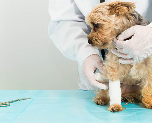
It’s day 18. The wound is clean, it’s moist, it’s covered. And it looks exactly the same as it did at day 10. No new granulation islands. No edge contraction. The exudate has shifted from serous to something slightly cloudier. The tissue around the margin is pale instead of the pink-red you’d expect in an actively healing wound.
This wound has stalled — and continuing the current protocol isn’t going to change that.
Chronic non-healing wounds are one of the most frustrating management problems in small animal practice. Not because they’re rare, but because standard wound care is genuinely appropriate for most wounds — and the moment when it stops being appropriate isn’t always obvious. Understanding what biologically derails the healing cascade, which patients are predictably at risk, and what a biologic intervention actually does differently is what separates a wound that closes in five weeks from one that doesn’t close in five months.
The Biology of the Stalled Wound: What Goes Wrong When Healing Stops
Normal wound healing follows a defined sequence: hemostasis, inflammation, proliferation, remodeling. Each phase has a beginning and an end. In a stalled wound, the sequence breaks down at the transition from inflammation to proliferation — and the wound gets stuck cycling through an inflammatory response that can’t resolve.
Three mechanisms drive most of what goes wrong.
Biofilm. Biofilm is present in approximately 60% of chronic wounds, compared to just 6% of acute wounds. That difference isn’t incidental — biofilm is both a consequence of chronicity and a driver of it. The extracellular polymeric substance (EPS) matrix that holds biofilm together creates three compounding barriers: a physical barrier that blocks keratinocyte and fibroblast migration; a metabolic barrier, generating hypoxic and acidic gradients that suppress cell energy production; and an immunological barrier that masks bacterial surface signals from host immune cells while bacterial toxins actively damage wound bed tissue. S. aureus α-toxin depletes ATP in keratinocytes. P. aeruginosa phospholipase causes direct necrosis. The biofilm doesn’t just prevent healing — it actively dismantles the wound bed.
MMP dysregulation. Matrix metalloproteinases are necessary for normal wound remodeling, but in chronic wounds, elevated MMP-9 creates a protease-dominant environment that degrades the extracellular matrix scaffolding before new tissue can form. Bacterial proteases compound this — S. aureus degrades immunoglobulins and complement proteins; certain anaerobes target type IV and V collagen directly. The wound bed loses its structural matrix faster than it can replace it.
Cell cycle arrest. Fibroblasts from chronic wounds exhibit growth arrest driven by elevated CDK inhibitors p21 and p27. These cells accumulate in the wound but can’t proliferate. Meanwhile, excess TGF-β — present in chronically inflamed wounds — imposes a secondary brake on keratinocytes via Smad2/3 phosphorylation, preventing the epithelial migration needed to close the wound surface. The cellular machinery is present but biochemically locked.
The result is a wound that looks stable but is doing nothing — consuming resources, resisting standard dressings, and waiting for a different signal.
Which Patients Get Stuck: Common Causes in Dogs and Cats
Certain wound types and patient profiles are predictably prone to chronicity. Recognizing them early changes the management calculus.
Post-surgical dehiscence. The most common scenario in general practice. A wound that should have closed primarily reopens — most often due to tension, infection, or patient interference — and then fails to granulate adequately over the following weeks. The surgical field has already been traumatized once; fibroblasts in the wound margins may be compromised, and secondary biofilm establishment is common. Revision surgery in a poorly vascularized, previously disrupted field carries its own risks. This is often where a biologic intervention has its clearest return.
Pressure ulcers in recumbent patients. Dogs with neurological or orthopedic conditions who are non-ambulatory develop pressure sores over bony prominences that are mechanically and metabolically difficult to heal. Sustained pressure impairs capillary perfusion, creating a hypoxic wound bed — precisely the environment that prevents normal cellular proliferation and is most permissive to biofilm. These wounds can persist for months to years under standard care.
Bite wounds and infected wounds in immunocompromised patients. FIV-positive cats, patients on chronic corticosteroids, and animals with concurrent systemic disease represent a high-risk group. A bite wound in a healthy cat is an acute problem with a predictable course. The same wound in an immunocompromised patient can fail to resolve for months, particularly when polymicrobial antibiotic-resistant biofilm establishes in the wound bed.
Patients with endocrine disease. Hyperadrenocorticism and diabetes mellitus are well-documented drivers of impaired wound healing in dogs. Elevated cortisol suppresses fibroblast function, impairs collagen synthesis, and blunts the immune response needed to clear early wound contamination. Any patient presenting with a chronically non-healing wound that doesn’t fit the wound’s timeline should be screened for underlying endocrinopathy — no biologic intervention fully compensates for uncontrolled Cushing’s disease.
The common thread across these patients is that their wound biology has been disrupted at a systemic or local level in a way that standard topical care cannot address.
How Amniotic Membrane Powder Restarts the Cascade — Step by Step
Standard wound care — appropriate moisture, debridement, infection control — creates the conditions for healing. What it doesn’t do is provide the biochemical signal the wound needs to move from inflammation to proliferation. That’s the gap a biologic like Healion fills.
Decellularized amniotic membrane powder delivers a preserved payload of growth factors that are absent or depleted in the chronic wound environment. Measured concentrations from amniotic membrane preparations include epidermal growth factor (EGF), fibroblast growth factor (FGF), hepatocyte growth factor (HGF), keratinocyte growth factor (KGF), nerve growth factor (NGF), and TGF-β — the same signals that coordinate normal wound healing but that chronic wounds can no longer generate at adequate levels.
Lyophilized amniotic membrane preparations show significantly higher bioactive factor concentrations than fresh tissue — up to 3.8x the collagen content, 5.2x the VEGF, and 6.8x the bFGF per milligram. That concentration matters clinically: a lyophilized powder applied to a chronic wound bed delivers a more concentrated signal than fresh membrane or simple moist dressings.
The mechanism at the cellular level is well-characterized. In chronic wounds, excess TGF-β locks keratinocytes in a growth-arrested state via Smad2/3 phosphorylation — the cells can’t migrate or proliferate. Amniotic membrane EGF activates MEK and attenuates Smad2/3 phosphorylation, releasing the TGF-β-imposed brake on keratinocyte proliferation. This isn’t blunt inflammation suppression — it’s modulation of a specific pathway that the chronic wound has dysregulated. Minimal TGF-β signaling is still necessary for proper migration; the AM restores balance rather than eliminating a signal.
Simultaneously, amniotic membrane shifts macrophage behavior from pro-inflammatory (M1) toward pro-remodeling (M2), elevating IL-10 (anti-inflammatory), supporting controlled TGF-β for appropriate matrix remodeling, and increasing MMP-1 and MMP-3 for organized ECM turnover. The net effect is a wound environment that can finally transition out of the inflammatory phase and begin building new tissue.
In a controlled study of lyophilized amniotic membrane in dogs with full-thickness wounds, closure rates at week 3 were 88.6% in the treatment group versus 55.7% in controls. By week 5, treatment wounds showed restored epidermis, remodeled dermis, and recovered hair follicles — structural markers of genuine tissue regeneration, not just wound closure. A separate AVMA study of an ECM wound dressing in Beagles showed significantly higher epithelialization percentages at days 7, 9, 12, and 18 compared to standard protocol, with better histologic repair scores.
Healion also retains antimicrobial activity from native amniotic tissue — β-defensins with documented activity against P. aeruginosa and S. aureus including MRSA strains. In a wound where biofilm is part of the problem, this is a clinically meaningful addition to the protocol.
Practical Decision Points: When Should a GP Vet Reach for a Biologic?
The clinical question isn’t whether amniotic membrane powder works — the evidence supports it. The question is when in the management timeline it becomes the right tool.
A reasonable framework: if a wound is not showing clear signs of progression by day 14 to 21 under appropriate standard care, treat that as a stall signal rather than normal variation. Specifically:
- No granulation islands forming at 14 days
- Wound edges not contracting
- Exudate shifting from serous to cloudy or purulent despite appropriate antibiotic coverage
- Tissue at the wound margin appearing pale or grey rather than pink-red
- Culture showing resistant organisms or polymicrobial contamination
At that point, consider whether there’s a systemic reason the wound is stalling — run a culture if you haven’t, screen for endocrine disease if the patient profile suggests it, reassess nutrition and pain management. Then layer in the biologic.
Healion’s bellow dispenser allows direct, sterile application to the wound bed without secondary processing — the powder format suits open wounds with irregular geometry and doesn’t require the wound to be prepared beyond standard debridement. It fits into an existing wound care protocol without requiring additional equipment or technique change.
The conversation with the client is straightforward: standard dressings keep the wound clean and provide the right environment. The powder provides the biological signal the wound has lost — growth factors and structural cues that tell the cells what to do next. That framing holds up to client questions and positions the intervention accurately.
For patients at high risk of chronicity — post-surgical dehiscence in an oncology patient, pressure sores in a recumbent dog, bite wounds in an immunocompromised cat — there’s a reasonable argument for starting the biologic earlier rather than waiting for the stall to establish.
Learn more about Healion’s mechanism and application at RethinkHealing.com.