
When Surgical Wounds Reopen in Dogs and Cats
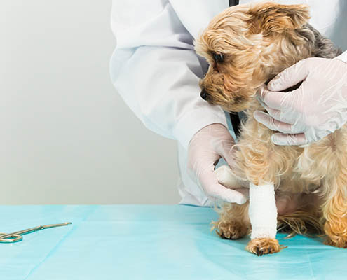
The surgery went well. The incision looked clean at discharge. Three days later the owner is back — the wound edges have separated, there’s discharge, and the tissue underneath doesn’t look like it’s planning to close on its own. This is post-surgical wound dehiscence, and it’s one of the more deflating events in general practice. The technical part of the case went right. The biology went wrong.
Understanding why surgical wounds fail — and what changes in the wound environment when they do — is the first step toward managing them more effectively. Amniotic wound matrix has a defined role in that recovery, but only if you know when to reach for it and why it works differently from standard wound care.
Why Surgical Wounds Sometimes Fail to Close
Surgical wound dehiscence isn’t one problem — it’s several different problems that produce the same clinical presentation. The mechanical failure mode and the biological failure mode require different interventions, and conflating them leads to the common mistake of simply re-closing a wound that isn’t ready to be closed again.
Mechanical dehiscence happens when suture material fails, knots slip, or the tissue itself tears through. It’s most common in locations under tension — perineal repairs, limb wounds near joints, closures over bony prominences — or when the patient interferes with the incision. In these cases the wound edges are usually viable, granulation is intact, and primary re-closure or tension-relieving techniques can work.
Biological dehiscence is different. It happens when the wound environment itself is hostile to healing — when inflammation is dysregulated, when local perfusion is compromised, or when the patient’s systemic disease is actively working against wound repair. In these cases, the tissue at the wound edges looks dull, pale, or frankly necrotic, re-approximation fails again, and the wound deteriorates in a predictable downward spiral.
The patients at highest risk for biological dehiscence are the ones already on your radar: dogs with hyperadrenocorticism, diabetic patients, animals on chronic immunosuppression, and patients with poor nutritional status. A 2025 case report in BMC Veterinary Research documented a dog with Cushing’s disease and confirmed impaired wound healing and heightened infection susceptibility — a combination that makes even routine incisions unpredictable. These are the cases where reaching for wound support early, before the wound declares itself, changes outcomes more than any secondary rescue intervention.
The Biology of a Disrupted Wound: What Changes After Dehiscence
An intact surgical wound moves through three overlapping phases: inflammation resolves, proliferation builds granulation tissue and new epithelium, and remodeling reorganizes collagen into durable scar. When the wound opens, that sequence doesn’t simply pause — it resets and frequently gets stuck.
Dehisced wounds almost uniformly stall in a chronic inflammatory state. Neutrophils and macrophages remain at elevated levels long past the point where they’re useful; matrix metalloproteinases (MMPs) are overexpressed and begin degrading the very growth factors and ECM components the wound needs to progress. Vascular endothelial growth factor (VEGF), basic fibroblast growth factor (bFGF), and the structural collagen scaffold that supports cell migration are all depleted faster than the wound can replace them.
This is where the wound biology of dehiscence diverges from the biology of a clean acute wound — and it’s why standard wound management (debridement, moisture balance, bandage changes) is necessary but often insufficient. Those interventions remove obstacles. They don’t replace what’s missing.
A 2025 study in Scientific Reports (Hussein et al.) provided direct mechanistic data in a canine full-thickness wound model comparing lyophilized amniotic membrane to saline controls. Lyophilized amniotic membrane retained significantly higher concentrations of VEGF (28.12 ± 7.6 ng/mg versus 5.43 ng/mg in fresh form) and bFGF (13.3 ± 6.89 ng/mg versus 1.97 ng/mg), along with collagen at 217.74 µg/mg versus 57.3 µg/mg. Dogs treated with lyophilized AM showed faster granulation tissue formation in the first week, better re-epithelialization, and superior collagen remodeling in subsequent stages compared to controls. The powder format concentrates and delivers exactly the growth factors and matrix proteins the chronic inflammatory environment has degraded.
Risk Factors That Predict Post-Surgical Wound Breakdown in Dogs and Cats
Not every dehiscence is preventable, but most cases that spiral into chronic wound management are predictable in advance. The risk factors cluster into three categories.
Concurrent endocrine and metabolic disease. Hyperadrenocorticism, hypothyroidism, and diabetes mellitus all impair wound healing through different mechanisms — cortisol excess suppresses fibroblast proliferation and collagen synthesis; thyroid deficiency slows all phases of repair; hyperglycemia impairs neutrophil function and creates an environment that favors biofilm formation. These patients need pre-surgical optimization where possible and early wound support when it isn’t.
Immunosuppression. Dogs on chronic corticosteroids, cyclosporine, or other immunosuppressants are at higher risk for both infection and impaired repair. The same mechanisms that suppress the inflammatory disease also suppress the productive inflammation that initiates wound healing. Post-surgical wound care protocols for these patients should anticipate the need for augmentation rather than waiting for failure.
Location, closure tension, and patient behavior. Perineal repairs, wounds over the distal limbs, and closures in high-motion areas are mechanically disadvantaged. Patients that lick, chew, or are difficult to confine compound the risk. A 2025 study of TPLO wound complications (Filippo et al., Vet Sci) highlighted that SSI rates in this common orthopedic procedure are clinically significant enough to drive antibiotic protocol debates — and TPLO wounds are in a high-motion location on a patient who is frequently non-compliant with restricted activity.
Large skin defects and feline patients. Cats present a specific challenge: their skin is relatively inelastic, their vascular supply to skin is more tenuous than in dogs, and their tolerance for bandaging is lower. A 2026 case report in BMC Veterinary Research (Waleed et al.) documented successful management of a feline skin defect covering 12–15% of body surface area following necrosis from subcutaneous fluid therapy. The wound was managed with amniotic membrane support and achieved complete epithelialization by day 33, with full hair regrowth and no scarring by day 74. The case is a practical illustration of what biological scaffold support enables in a wound that would otherwise require prolonged open wound management or skin grafting.
How Amniotic Wound Matrix Powder Restores the Healing Environment
The clinical decision point for Healion in post-surgical wound management isn’t the perfectly clean wound that just needs time. It’s the wound that isn’t behaving — the one that’s stalled, inflamed, producing more exudate than expected, or beginning to show surface necrosis at day five or six when it should be progressing.
Healion is a decellularized amniotic membrane powder that delivers the extracellular matrix proteins, growth factors, and cytokines that a disrupted wound has lost. Because it’s fully decellularized, it carries no risk of immune reaction or disease transmission. Because it’s delivered as a powder via the bellow dispenser, it reaches irregular wound geometry that liquid or sheet products can’t fill evenly — debrided pocket wounds, sutured dehiscences with partial opening, and post-surgical wounds with uneven surfaces all benefit from the powder format’s ability to coat and adhere to the entire wound bed.
The mechanism isn’t replacing the wound’s biology — it’s restoring the substrate that allows normal biology to proceed. Healion modulates the inflammatory environment, delivers the growth factors (VEGF, bFGF) that drive angiogenesis and fibroblast migration, and provides the ECM scaffolding that organizes new collagen deposition. The result isn’t just wound closure — it’s wound closure with reduced scarring and better tissue architecture, which matters for functional outcomes in high-motion locations.
A 2026 case report (Rodríguez-Aguilar et al., Case Reports in Veterinary Medicine) documented a post-nodulectomy wound in a dog managed with multimodal biologic wound support using the TIME wound-bed preparation framework. The outcome showed no dehiscence, no recurrent necrosis, and no systemic complications through the full follow-up period — the kind of result that becomes the expectation rather than the exception when wound support is built into the protocol from the start, not added after the first failure.
For cases where wound dehiscence is a real possibility — endocrine disease, immunosuppression, high-tension closures, feline skin defects — having Healion as part of your post-surgical protocol from the first bandage change is the intervention that prevents the downward spiral. By the time a wound is visibly failing, you’ve already lost days of healing potential that early support could have preserved.
Learn more about Healion amniotic wound matrix at RethinkHealing.com.
Sources
- Hussein KH, et al. Lyophilized amniotic membrane in canine full-thickness wound healing: growth factor retention and histopathologic outcomes. Sci Rep. 2025. PMID: 40320419.
- Waleed A, et al. Management of a large feline skin defect with amniotic membrane: complete epithelialization and hair regrowth. BMC Vet Res. 2026. PMID: 41877121.
- Rodríguez-Aguilar SE, et al. Post-nodulectomy wound management with biologic support using TIME framework in a dog. Case Rep Vet Med. 2026. PMID: 42707463.
- Moon JS, et al. Hyperadrenocorticism and impaired wound healing in dogs. BMC Vet Res. 2025. PMID: 41053838.
- Filippo LD, et al. Surgical site infection rates in TPLO procedures — perioperative antibiotic protocols. Vet Sci. 2025. PMID: 40267006.