The Use of BioSIS in Hernia Repair
While internal obturator muscle transposition (IOT) has, for a while now, been regarded as the primary surgical treatment for perineal hernia repair in dogs, recent studies have shown porcine small intestinal submucosa (SIS) may perform better.
What Is Herniorrhaphy?
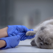
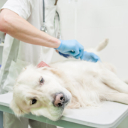
Another term for hernia repair that you may have heard is herniorrhaphy. A perineal hernia occurs when the pelvic diaphragm muscles are incapable of supporting the pelvic organs because they are too weak. Causes may include chronic constipation, tenesmus, and weakening of the levator ani muscle. The condition commonly occurs in middle-aged to older, intact male dogs.
SIS vs. IOT
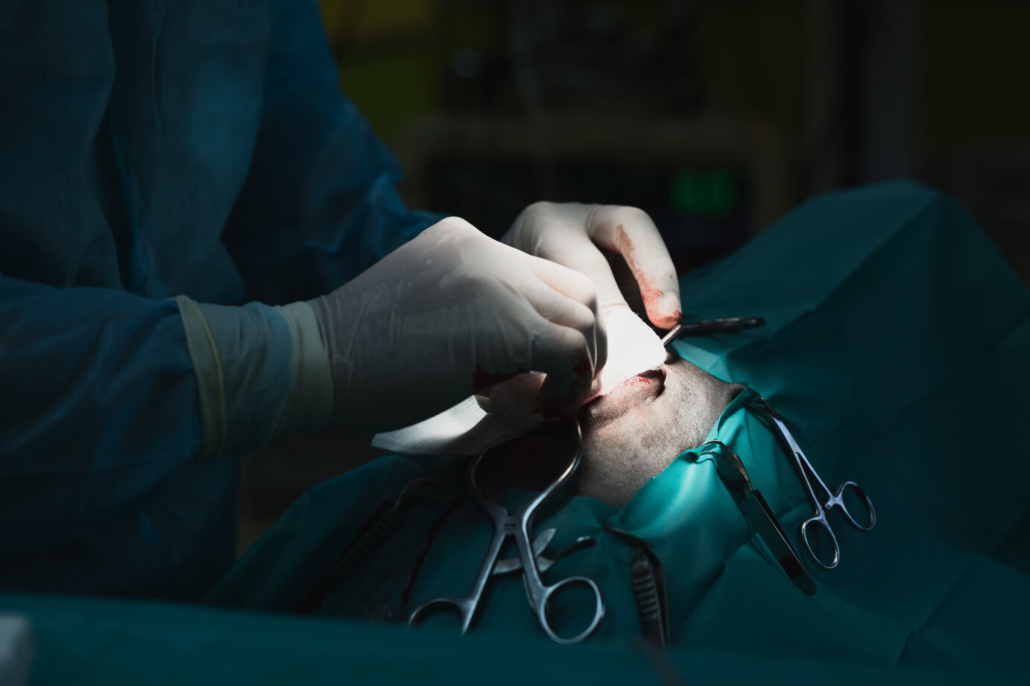
When using small intestinal submucosa (SIS) as a biomaterial to treat hernia repair on dog subjects, investigators used a 4-ply SIS sheet trimmed to dimensions slightly larger than the pelvic diaphragm defect, rehydrated it using sterile saline, and secured it to the muscles using sutures. SIS material promotes blood vessel and tissue ingrowth that is structurally similar to host tissues.
It meets all the recommendations of an ideal hernia repair tissue such as inexpensive, resistant to infection, no inflammatory response, and inhibits adhesion and fistula formation. It’s superior to other substrates in that it has shown the least amount of residual implant material and lower infection rates.
The SIS technique is purported by investigators to have less potential for complications and is easier to perform over the IOT technique. It also has been proved to have the same biomechanical strength and stiffness as the IOT technique.
Why Is SIS Better for Hernia Repair?
Taken from the jejunum of pigs, porcine SIS is an acellular extracellular matrix. It is made up of type I collagen and contains vascular endothelial growth factor and fibroblast growth factor. This biomaterial is also known to be used as a xenograft for vascular grafts, Achilles’ tendon repair, urinary bladder augmentation, and dura mater grafts among others.
In experiments on dogs, both small intestinal submucosa and internal obturator transposition resulted in a band of fibrous connective tissue, but on IOT patients, the band was objectively wider. IOT repairs also regularly developed the following issues: multifocal and randomly scattered aggregates of lymphocytes within the fibrous bands, shrunken and eosinophilic clusters and individual myofibers, necrosis, and small foci of mineralization . SIS repairs showed no signs of aggregates of lymphocytes, necrosis, or inflammation.
The internal obturator transposition has generally been preferred over other surgical techniques for perineal hernia treatment like superficial gluteal muscle transposition, semitendinosus muscle flap, porcine dermal collagen implants, and synthetic mesh implants. However, with new research and findings, it looks like that has now changed to small intestinal submucosa.
If you’d like more information on this topic, contact us today. We’d love to hear how you may be (or already are) able to incorporate this technique into your practice to help your patients. And for more industry-leading science and medicine blogs for veterinarians, check out our main blog page.