polypropylene meshAccording to the Veterinary Society of Surgical Oncology, spinal tumors in dogs are uncommon, but when they do happen, 90% of spinal tumors occur in large-breed dogs. Meningioma is the most common and presents most often in the cervical spinal cord. Regardless of the type of spinal tumor a veterinarian may face treating at some point in their career, it’s always best to be prepared. Recommending the best treatment plan for a dog with a spinal tumor diagnosis is critical, as spinal tumors are slow-growing and may need more time to respond to treatment.

Signs and Symptoms of Spinal Tumors

Signs and symptoms of spinal tumors that dog owners may notice that require an immediate examination by a veterinarian include:

  • Changes in movement
  • Changes in coordination
  • Limb weakness
  • Neurological changes
  • Pain

These signs can include lethargy, difficulty getting up and down, dragging limbs or limping, depression, decreased appetite, or difficulty going potty. If a dog presents with any of the above symptoms, a vet may order some diagnostic tests for spinal tumors, such as:

  • CT Scan
  • MRI
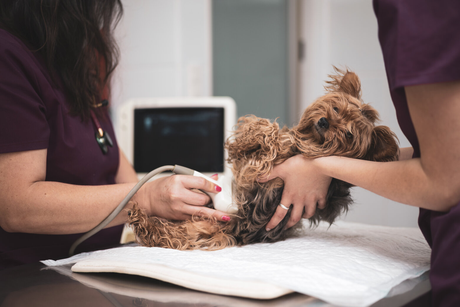
  • Ultrasound
  • Chest X-ray
  • Bloodwork
  • Urinalysis
  • Biopsy

Spinal Tumor Treatment Options

A spinal tumor diagnosis can be frightening, but many treatment options are available for dogs today. The tumor type, location, and grade determine the best course of treatment, but these are the most common ways to help dogs with a spinal tumor diagnosis:

Surgery

Surgery for a spinal tumor can be complicated based on the tumor’s location. However, regarding veterinary neurologist technologies, Vetrix is an industry leader. Vetrix BioSIS for neurology is a platform technology that can be used for several surgical applications, including as a dural graft for spinal surgeries and spinal tumor surgeries. When used for these applications, the contact between BioSIS and the surrounding tissue allows cells to migrate, separate, and differentiate within the bio-scaffold. This matrix is easy to handle and simple to use but strong enough to hold sutures and support weakened tissue. If the tumor can be removed without impacting the functionality of the spinal cord, surgery is an excellent treatment option.

Chemotherapy

Chemo treats spinal tumors in dogs that have already spread or are at high risk for spread. Veterinarians make specific and informed recommendations based on the tumor type.

Radiation Therapy

Radiation therapy may be used alone or with surgery as part of a dog’s spinal tumor treatment plan. Again, this recommendation will be specific to a dog’s tumor type.

Palliative Therapy

Palliative therapy includes things like antibiotics and painkillers that help maintain a dog’s quality of life but does nothing to slow the progression of the spinal tumor. Palliative therapy is meant to keep a dog comfortable when no other treatment options are available or have been exhausted.

Spinal Tumor Prognosis

As with any cancer, the prognosis varies by case, type, and tumor location. The earlier the cancer is diagnosed and treated, the better the chances treatment will be successful. It also helps to have the best veterinary science technology and tools at your fingertips. 

For more information on any Vetrix technologies, contact us with your questions.

ultrasound diagnostics in caninesYour furbaby needs gastrointestinal surgery, and you have all kinds of questions. We’re here to discuss how you can prepare and what you need to know.
What Is Gastrointestinal Surgery?

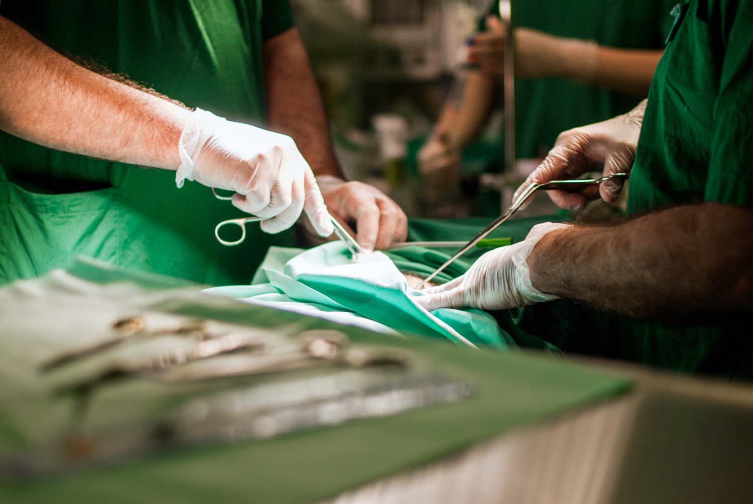
Gastrointestinal surgery is an umbrella term for any surgery involving the stomach or intestines. There are numerous reasons a pet may need gastrointestinal surgery, ranging from blockages and biopsies to twisting stomachs or tumor removals. The exact nature of the gastrointestinal surgery depends on the animal’s individual needs and whether the surgery is planned or performed as an emergency.

How Much Does Gastrointestinal Surgery Cost?

Again, it varies. The cost of gastrointestinal surgery will depend on the following:

  • Type of surgery performed
  • Size of the pet
  • Severity of the condition
  • Time your pet needs to be hospitalized before and after surgery

On average, pet parents should expect a bill anywhere from $2,000 to $10,000. To help you balance the care and cost of gastrointestinal surgery, ask your veterinary team for a cost estimate before the procedure. 

Balancing Care and Cost for Vets and Pet Owners

A veterinary surgeon’s primary concern when a pet needs gastrointestinal surgery isn’t money. It’s helping the patient get better and live a comfortable, happy life. Be bold and ask questions or express concerns. While we must submit payment for services rendered, an excellent veterinary office is committed to:

  • Explaining any gastrointestinal procedure your pet may need
  • The process and what to expect
  • Providing a cost estimate
  • Follow-up and at-home care

Many veterinary offices will work with you to establish a payment plan if necessary. They can also often provide information about pet insurance that will likely cover some gastrointestinal surgery costs. In addition to working out the cost (and payment plan if needed), your vet should also work with you to ensure you understand aftercare instructions while your pet is recovering.

Some things your vet may tell you to do after your pet has had gastrointestinal surgery and finished its hospital stay include:

  • Monitor your pet closely and keep them calm to prevent tearing of sutures
  • Keep your pet’s cone on so they can’t lick or chew their incision as it heals
  • Feed your pet small amounts of bland food and plenty of water as they slowly transition back to their regular diet
  • Administer pain medication as instructed

Gastrointestinal surgeries are often a lifesaving intervention. As a patient, working with a vet dedicated to balancing care and cost makes a big difference in delivering as positive and stress-free of an experience as possible. As a vet, the ability to confidently deliver as positive of an experience as possible relies on your dedication to staying up-to-date on the medicine and technology available to you to help you perform your job to the best of your ability. 

For more information on gastrointestinal problems, surgery, and solutions (for both pet parents and veterinarians) we think you’d be interested in reading more here: How Regenerative Medicine Can Help with Gastrointestinal Problems and Surgery.

*Note: The advice provided in this post is intended for informational purposes and does not constitute medical advice regarding pets. Please make an appointment with your vet for an accurate diagnosis, treatment plan, and cost assessment of your pet’s condition.

Veterinary Neurology Vetrix

When a patient has a wound that’s too big to close on its own or a wound that becomes chronic and doesn’t heal by itself, treatment with a skin graft may be beneficial in obtaining wound closure. Skin grafts help quickly and effectively restore function and provide better cosmetic results than other treatment options. Additionally, skin grafts protect from infections and parasites. The Vetrix BioSIS Technology we provide is a regenerative medicine beneficial in remodeling, regrowing, and repairing wounds encountered in the veterinary field.

Two Common Skin Graft Procedures in Veterinary Medicine

When it comes to common household pets, veterinarians are often most likely to perform one of the following types of skin grafts when necessary for wound treatment:

  • Total Skin Graft (or Full-thickness skin graft): This skin graft includes the epidermis and dermis. It involves removing a piece of skin and the fat from the underside of the skin. This grafting procedure requires the donor site to have enough surrounding loose skin so the incision can be closed.
  • Partial Thickness Skin Graft (or Split-thickness skin graft): This skin graft involves shaving a thin layer of skin, 0.2 to 0.4mm, off the donor site. The cutting plane of this graft remains above the hair follicles so that no hair will grow from this skin graft. The donor site will heal independently and doesn’t require closure, but it may be more painful than a total skin graft because the exposed nerve endings will need time to heal. It’s typically used in burn victims with limited normal skin for grafting.

Veterinarians will guide their patients on the best skin graft option for their particular wound.

Treating Skin Grafts With Regenerative Veterinary Medicine

Once it’s been determined that a pet needs a skin graft to heal a wound, consider a regenerative medicine solution as part of your treatment. Unlike synthetic materials or other biological grafts, Vetrix Technology provides a structure for healthy tissue to grow across and incorporate into the extracellular matrix, resulting in wholly remodeled, vital, and fully vascularized tissues. During the healing process, Vetrix BioSIS is replaced with the body’s native tissue, developing into a permanent repair without the long-term presence of a foreign body. After healing is complete, patients are left with healthy, natural tissue.

BioSIS is a great regenerative medicine solution for skin grafts because it facilitates angiogenesis and rapid remodeling, allowing the body’s defense mechanisms to react and respond to any potential infection. It promotes safe and sterile healing of the wound.

Download the BioSIS Informational Brochure to learn more about this technology and how it can aid in healing skin graft patients.

Get the brochure.

Caring for and treating patients with chronic wounds can sometimes be frustrating and discouraging. You so desperately want to give them the best wound care possible and see them live their lives to the fullest, but nothing seems to offer a permanent solution. You may be able to improve your results with the BioSIS method.

Using Porcine Small Intestinal Submucosa (SIS) for Wound Care

Researchers say that using porcine SIS to treat chronic wounds has improved healing time and permanency. Due to its makeup, an acellular, biological extracellular matrix (ECM), porcine small intestinal submucosa draws in the host’s cells to attach and multiply. This aids in quicker, more permanent healing.

Growth compounds like collagen, elastin, glycosaminoglycans, and proteoglycans help with the healing process. And it’s not just one cell that’s attracted to the matrix. Porcine SIS attracts numerous cell types for a healthy, diverse layer formation. Since it takes more than growth factors to aid in the healing process, it’s fortunate that SIS has been found to decrease matrix metalloproteinases (MMP) activity. These endogenous proteolytic enzyme levels usually rise with chronic wounds.

Even better, the mechanical properties of multi-layer SIS are stronger and degrade slower as opposed to single-layer SIS. Pertaining to the repair of different kinds of wound types, these two SIS products provide flexibility when choosing a biologically-active ECM.

1-Layer VS. 3-Layer SIS

When choosing between 1-layer SIS and 3-layer SIS, studies show a thicker matrix may be preferable. One study done in diabetic mice showed the 3-layer SIS didn’t require application as frequently as the 1-layer matrix. Where both SIS layers were applied to full-thickness wounds on the day of wounding and three days later, the thicker matrix showed larger sections of unincorporated layers—meaning there was still significant space in the matrix for cells to mitigate.

This is good to know for patients that may have owners who live farther away and travel a greater distance for appointments or cannot afford frequent applications.

More Reasons to Consider the SIS Method

Besides reducing MMP levels, you may also want to consider the SIS method for wound care because it contains proteins that foster cell attachment and growth factor binding sites, sequester matrix-degrading enzymes, and enhance cellular filtration into damaged tissue. In addition to these positive effects, it also supports new blood vessel growth, which is vital to wound recovery.

Overall, small intestinal submucosa has been found to not only increase healing in wounds but also the rate at which the wounds recover compared to the standard of wound care.
If you’d like to further discuss this method of wound care or have any questions, please contact us. We would be happy to speak with you! And if you’d like to read more on topics like this, visit our blog page.

BioSIS is revolutionizing the way veterinarians are able to care for seriously wounded patients. Due to its framework and ability to attract cells, BioSIS may shorten healing time and ensure organized tissue regrowth in the damaged area. Good blood flow is key to using the BioSIS method successfully. Read on to discover how to get the best results when using this method of wound care in your patients.

Use the Horizontal Mattress Technique

Proper blood flow is essential to successfully using BioSIS as a biological scaffold. Using the horizontal mattress technique to fixate the graft encourages blood flow to the wound site, by connecting to many points of blood supply. This also enables you to slide the edges of the implant under the undermined tissue edges and allows for further connectivity to the patient’s healthy blood supply.

Throughout BioSIS insertion, you can also use “tacking” to apply independent sutures and ensure it’s secure and flush against the wound.

Perforate the BioSIS Scaffold if Necessary

To avoid inflammation, fenestration may be necessary. This usually only applies to larger wounds to keep cells and other fluids from accumulating between the BioSIS scaffold and healing flesh. It is also helpful in preventing a hematoma from forming.

One major threat to the success of this procedure is dried blood. This generates inflammation, which is crucial to limit throughout the healing process. Stoppage of blood flow can prevent cells from evenly attaching to the scaffold and deter the healing process.

Keep the Matrix in Place

Once cells begin to lay their own collagen matrix, it’s important to be extremely careful when changing bandages. As the patient’s own cells take over the BioSIS graft, it will be resorbed at different rates—not all at once. When changing bandages, it’s imperative that as much of the extracellular matrix from the implant be left intact to continue healing. Using a non-adherent bandage to dress the wound will help you to change it more easily.

Within the first 6-10 days of post-operative recovery, the wound will turn caramel-colored as the damaged tissue is replaced and repaired. Instead of thoroughly cleaning the wound and scraping off this pigment, simply irrigate with saline and apply another non-adherent bandage. This way, all of the graft is resorbed and healing is even across the entire contusion.

Allow/Encourage Wound Site Mobility

When bandaging the patient, allow for as much mobility as possible while still covering the entirety of the wound site. Mobility encourages blood flow which, as we mentioned earlier, is essential to the success of this method of healing.

Based on extensive research, we’ve found that mobility increases the rate at which the healing occurs and makes for a better off healing process overall. This is because the cells attaching themselves to the scaffold receive their direction from the surrounding matrix’s blood flow. If blood flow is weak or immobile, the cells will have no direction to follow. Just remember, form follows function.
We’d love to answer any questions you may have about this technique. Contact us to learn more or tell us how you’ve been able to help your patients using BioSIS technology. You can also visit here for more blogs on topics like this.

While internal obturator muscle transposition (IOT) has, for a while now, been regarded as the primary surgical treatment for perineal hernia repair in dogs, recent studies have shown porcine small intestinal submucosa (SIS) may perform better.

What Is Herniorrhaphy?

Another term for hernia repair that you may have heard is herniorrhaphy. A perineal hernia occurs when the pelvic diaphragm muscles are incapable of supporting the pelvic organs because they are too weak. Causes may include chronic constipation, tenesmus, and weakening of the levator ani muscle. The condition commonly occurs in middle-aged to older, intact male dogs.

SIS vs. IOT

When using small intestinal submucosa (SIS) as a biomaterial to treat hernia repair on dog subjects, investigators used a 4-ply SIS sheet trimmed to dimensions slightly larger than the pelvic diaphragm defect, rehydrated it using sterile saline, and secured it to the muscles using sutures. SIS material promotes blood vessel and tissue ingrowth that is structurally similar to host tissues.

It meets all the recommendations of an ideal hernia repair tissue such as inexpensive, resistant to infection, no inflammatory response, and inhibits adhesion and fistula formation. It’s superior to other substrates in that it has shown the least amount of residual implant material and lower infection rates.

The SIS technique is purported by investigators to have less potential for complications and is easier to perform over the IOT technique. It also has been proved to have the same biomechanical strength and stiffness as the IOT technique.

Why Is SIS Better for Hernia Repair? 

Taken from the jejunum of pigs, porcine SIS is an acellular extracellular matrix. It is made up of type I collagen and contains vascular endothelial growth factor and fibroblast growth factor. This biomaterial is also known to be used as a xenograft for vascular grafts, Achilles’ tendon repair, urinary bladder augmentation, and dura mater grafts among others.

In experiments on dogs, both small intestinal submucosa and internal obturator transposition resulted in a band of fibrous connective tissue, but on IOT patients, the band was objectively wider. IOT repairs also regularly developed the following issues: multifocal and randomly scattered aggregates of lymphocytes within the fibrous bands, shrunken and eosinophilic clusters and individual myofibers, necrosis, and small foci of mineralization . SIS repairs showed no signs of aggregates of lymphocytes, necrosis, or inflammation.

The internal obturator transposition has generally been preferred over other surgical techniques for perineal hernia treatment like superficial gluteal muscle transposition, semitendinosus muscle flap, porcine dermal collagen implants, and synthetic mesh implants. However, with new research and findings, it looks like that has now changed to small intestinal submucosa.

If you’d like more information on this topic, contact us today. We’d love to hear how you may be (or already are) able to incorporate this technique into your practice to help your patients. And for more industry-leading science and medicine blogs for veterinarians, check out our main blog page.

Boxers hold a special place in the hearts of many dog lovers. Atlas, a rescue from Florida, is one of them. Atlas was in line for an amputation due to a large sarcoma mass tumor that had grown on his leg. Dr. Steven Whittle, who worked with the Atlanta Boxer Rescue on special cases, heard about Atlas and volunteered to help. 

Dr. Whittle could see that the Boxer’s situation wasn’t good, with Atlas facing either an amputation or euthanasia. Still, he wasn’t convinced those were the only solutions available for the friendly pup. He chose a more optimistic approach, introducing BioSIS from Vetrix to the treatment plan. 

Giving Atlas a Fighting Chance 

Dr. Whittle felt certain advanced treatments would give Atlas a better chance to live a long and happy life. Having used BioSIS successfully to help many other patients in his veterinary practice, he had the knowledge he needed to make the right decision for Atlas’s surgery. Dr. Whittle felt positive knowing BioSIS would support the tissues in Atlas’s leg once the tumor was removed, providing regeneration for healthy healing.

What Makes BioSIS from Vetrix Different?

Vetrix BioSIS supports tissue repair, resulting in a remodeled, functional tissue without any premature breakdown of the material, calcification, or tissue erosion that can occur by synthetic materials. 

• BioSIS acts as a scaffold where the patient’s cells migrate and integrate. Over time, cells lay down their own collagen, which matures to form strong, permanent tissue repair. 

• BioSis provides permanent healing and long-term strength, while also resisting infection through the body’s own defense mechanisms.

Surgery Using BioSIS Begins

Atlas’s surgery was one of complexity. The team worked with Dr. Whittle to remove what had become a giant tumor for the mid-size dog. With a successful separation of the mass, Dr. Whittle began his work using Vetrix BioSIS ECM Technology. Applying two high-tech skin grafts, he was hopeful the regenerative treatment would work its magic to heal Atlas’s leg with no complications.

BioSIS from Vetrix Heals Again

It was no surprise to the veterinary team to see Atlas thrive as he healed from his BioSIS surgery. Yet no one was happier than Atlas’s adopted owner, forever thankful for Dr. Whittle’s insight to use BioSIS to save his dog’s life for six more years of quality living.

Learn More About BioSIS from Vetrix

Vetrix regenerative medicine, including BioSIS, rethinks healing to deliver innovative treatments to overcome illness, surgery, wounds, and eye conditions. Learn why veterinarians around the world count on Vetrix for exceptional patient care. Contact us today.